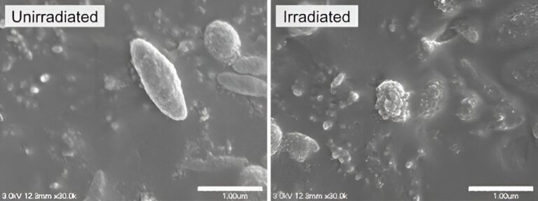
Left: Non-irradiated melanosomes show a smooth surface. Right: Irradiation by a picosecond laser with a pulse width of 450 ps at a wavelength of 1,064 nm and a flux of 8.50 J/cm² leads to melanosomes with their surface structure disrupted. Credit: Osaka Metropolitan University
Many people who are bothered by skin imperfections may turn to laser treatment. To improve the effectiveness and reduce complications from such laser therapy, a research team led by Osaka Metropolitan University developed an index of the threshold energy density, known as flux, and the dependent wavelength for picosecond lasers. The project was published on Lasers in Surgery and Medicine.
Picosecond lasers have been used in recent years to remove pigmented lesions. These lasers emit beams of energy in pulses lasting about a trillionth of a second. The lasers target the melanosomes, which produce, store and transport the melanin responsible for the pigment.
Postdoctoral fellow Yu Shimojo of the OMU Graduate School of Medicine and specially appointed professor Toshiyuki Ozawa and professor Daisuke Tsuruta of the school’s Department of Dermatology were among the researchers who developed this first picosecond laser marker for each of the wavelengths used in clinical practice in the treatment of pigs.
By comparing previously reported clinical studies, the researchers confirmed that clinical results showing low complication rates and high efficacy can be explained on the basis of these wavelength-dependent markers.
“The use of this marker is expected to play an important role in determining radiation conditions in clinical practice,” said Postdoc Fellow Shimojo. “Furthermore, applying picosecond laser therapy based on scientific evidence, rather than relying solely on the experience of physicians, is expected to improve the safety and efficacy of treatment.”
More information:
Yu Shimojo et al, Wavelength-dependent threshold fluxes for melanosome disruption for the evaluation of 532-, 730-, 755-, 785-, and 1064-nm picosecond laser treatment of pigmented lesions, Lasers in Surgery and Medicine (2024). DOI: 10.1002/lsm.23773
Reference: Spot laser treatment for skin blemishes gets clearer with new index (2024 March 27) Retrieved April 6, 2024 from
This document is subject to copyright. Except for any fair dealing for purposes of private study or research, no part may be reproduced without written permission. Content is provided for informational purposes only.


