June 25, 2024
By combining the laboratory infrastructures at BAM and HZB, more accurate measurements can be made
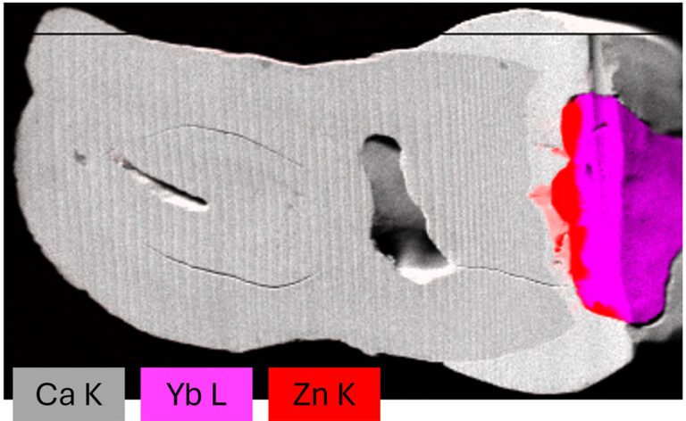
Composite micro-XRF image of Ca (white/tooth), Yb (magenta/filler) and Zn (red/sealant) distribution in a treated human tooth showing the diffusion of Zn from the sealant into the tooth. © Leona Bauer (TU Berlin/HZB)
A research team led by Dr. Ioanna Madouvalou has developed a method to more accurately depict the elemental distributions in dental materials than was possible in the past. The used confocal micro-X-ray fluorescence (micro-XRF) analysis provides three-dimensional elemental images containing distortions. These distortions occur when X-rays pass through materials of different density and composition. Using micro-CT data, which provide detailed 3D images of material structure, and chemical information from X-ray absorption spectroscopy (XAS) experiments performed in the laboratory (BLiX, TU Berlin) and at the BESSY II synchrotron light source, the researchers have improved the method.
“We can now make more precise measurements,” says Ioanna Madouvalou. “Absorption correction with micro-CT and XAS takes into account how strongly different materials absorb X-rays.” This was made possible through a combination of laboratory infrastructure at the BAM (Federal Institute for Materials Research and Testing) and the HZB SyncLab in conjunction with the BESSY II synchrotron light source. BESSY II provided tunable X-rays over a wide energy range (200 eV to 32 keV) necessary for detailed compositional analysis. Micro-CT and confocal micro-XRF investigations were then facilitated using laboratory setups using X-ray tubes as sources.
One of the materials investigated by Madouvalou’s team is dentin — a mineralized tissue that makes up most of the tooth, lies beneath the enamel, and plays a key role in transmitting sensations such as cold and heat. Its analysis is important in dentistry because, with dental fillings, elements often diffuse from the filling material into the dentin. “Our results enable detailed studies of such diffusion processes,” says Leona Bauer, PhD student at HZB and TU Berlin and first author of the study. They are important for improving the durability and biocompatibility of dental fillings and reducing the risk of secondary caries and other dental problems.
In addition to the investigation of materials for dentistry, the method offers applications in other fields where accurate 3D element distributions are required. These include the analysis of biological tissues, the investigation of catalytic materials, and the study of materials in environmental science. The flexibility of the measurement method could therefore have a positive impact on various research fields.
Publication:
Absorption correction for 3D elemental distributions of dental composites using laboratory confocal X-ray fluorescence spectroscopy
Leona J. Bauer, Frank Wieder, Vinh Truong, Frank Förste, Yannick Wagener, Adrian Jonas, Sebastian Praetz, Christopher Schlesiger, Andreas Kupsch, Bernd R. Müller, Birgit Kanngießer, Paul Zaslansky and Ioanna Madouvalou.
Analytical Chemistry (2024). DOI: 10.1021/acs.analchem.4c00116
Contact:
Helmholtz-Zentrum Berlin für Materialien und Energie
Dr. Ioanna Madouvalou
SyncLab Research Group – Combined X-ray Methods at BLiX and BESSY II
ioanna.mantouvalou(at)helmholtz-berlin.de
HZB Press Release, 24 June 2024


