OSAKA, Japan, April 4, 2024 — Laser therapy has gained popularity for treating skin blemishes. To improve the effectiveness and reduce complications from laser therapy, a research team led by Osaka Metropolitan University developed an energy density or flux threshold index for melanosome disruption, corresponding to the dependent wavelengths of picosecond lasers (ps) used for treatment. . The wavelength-specific radiation index will help clinicians determine the optimal endpoint for the treatment of melanocytic lesions based on numerical indices.
Picosecond lasers remove damage from the skin by delivering beams of energy in pulses lasting about a trillionth of a second. The lasers target the melanosomes, which produce, store and transport the melanin that is responsible for the pigment.
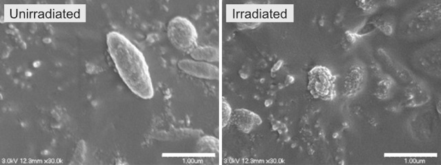
Scanning electron micrographs of melanosomes. Non-irradiated melanosomes show a smooth surface (left). Irradiation from a picosecond (ps) laser with a pulse width of 450 ps at a wavelength of 1064 nm and a flux of 8.50 J/cm² results in disruption of the surface structure of melanosomes (correctly). Courtesy of Osaka Metropolitan University.
In the experiment, the researchers irradiated a solution of melanosomes extracted from pig eyes using a ps laser with varying fluence. They measured the average particle size of irradiated melanosomes by dynamic light scattering. To observe melanosome disruption, they used scanning electron microscopy to determine disruption thresholds.
They developed a mathematical model and combined the model with each threshold taken and Monte Carlo light transfer to calculate the radiation parameters required to disrupt melanosomes within the skin tissue at various thresholds.
The researchers quantified radiation parameters for 532-, 730-, 755-, 785-, and 1064 nm ps laser treatments and determined wavelength-dependent thresholds for melanosome disruption. They determined the boundary fluxes to be 0.95, 2.25, 2.75 and 6.50 J/cm²respectively.
Assessment of radiation parameters from a threshold-based analysis provided numerical indices to define clinical endpoints for ps laser therapy. The markers quantitatively revealed the relationship between radiation wavelength, incident flux, and spot size required to disrupt melanosomes distributed at different depths in the skin tissue.
When the researchers compared these results with previously reported clinical studies, they were able to confirm that the calculated radiation parameters were consistent with clinical parameters that showed high efficacy and low incidence of complications.
Wavelength-dependent numerical markers for melanosome disruption could contribute to the effective evaluation and treatment of pigmented lesions.
“Using this index is expected to play an important role in regulating radiation conditions in clinical practice,” said researcher Yu Shimojo. “Furthermore, applying picosecond laser therapy based on scientific evidence, rather than relying solely on the experience of physicians, is expected to improve the safety and efficacy of treatment.”
According to the team, this is the first ps laser marker created for each of the wavelengths used in clinical practice to treat pigmented lesions. Although the thresholds for a 755 nm ps laser were previously determined, the wavelength dependence has not been investigated until now.
The research was published in Lasers in Surgery and Medicine (www.doi.org/10.1002/lsm.23773).


