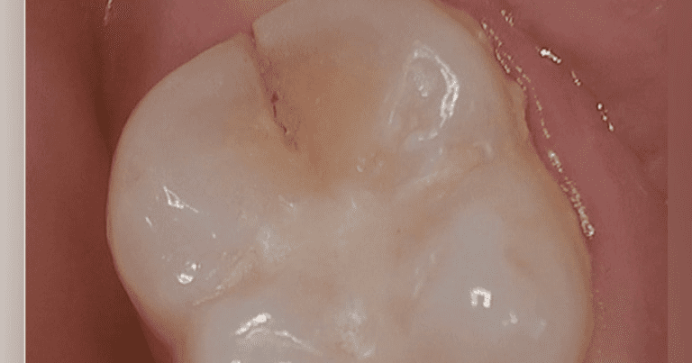
In our last article, “Dental Insurance Claim Reimbursement: Clinically Optimizing Scaling and Root Planing,” we discussed the non-negotiable claim for scaling and root planing reimbursement: visible bone loss. However, SRP codes are not the only CDT codes that are notoriously difficult to obtain reimbursement for dental insurance benefits. Indirect restorations—especially crowns and inlays (if the policy covers onlay treatment) and the restorative base of a core build-up—seem to provide just as much resistance. Are you like most providers and patients who believe that insurance companies just don’t want to pay benefits for claims submitted? Well, that’s not true.
Further reading… Dental Insurance Claim Reimbursement: Clinical Optimization of Scaling and Root Planing
Adjudication of claims
As I have spent time with various payers and their dentists, the majority of dental insurance adjusters want to “automatically” adjudicate claims. What does this mean? In most cases, if a claim is made for a crown replacement and the frequency restriction is not met, insurers want to allow this treatment as long as their submission requirements are met—that is, a periapical radiograph without periapical pathology and a narrative documenting the need. (Individual requirements vary.)
In my many claims experiences while in private practice, I have found that third party payers have been more than willing to return insurance benefits for treatment they should have received, often with less resistance than I thought there would be. . But what does it mean as a service that “must” benefit? Insurance companies have established their corporate policies, which serve as guidelines for their review team to compare when making adjudication recommendations (eg, “Was this treatment medically necessary at the time it was administered?”). This is not intended to judge your treatment recommendation, but to provide standardization.
Let’s look at specific requirements and criteria that are commonly checked for Crown claims. These are largely consistent across most insurance carriers, although it may vary slightly from payer to payer.
Indirect restorations: Crowns and inlays
Preoperative radiography is generally required for indirect restorations and restoration foundation codes. Note that the requirement is a radiographic image, not an intraoral photographic image. Some agencies require that this preoperative radiograph be periapical, and some require a periapical radiograph that captures the entire apex to facilitate complete radiographic evaluation of the tooth apex and surrounding tissues.
The clinical claims examiner tries to confirm that the apical area is free of pathology and treated appropriately, which can be documented with a final endodontic radiograph for greater success or, at the very least, in the narrative. Since capturing the apex does not give us a complete snapshot of the tooth’s health, the clinical crown must be evaluated, and this is done either by capturing the coronal structure in the same PA or by providing additional radiographs, more PAs or, more frequently , a bite.
Sometimes, practices provide intraoperative photos and/or x-rays, but these do not match preoperative radiography requirements. A common scenario of confusion, resulting in a request for more information, a negative decision such as denial or appeal, is a case involving a remote crown. The PA acquired before recording a new display or scan is preoperatively and should be shared in a narrative. During my peer conversations, offices would tell me, “The claim form says the previous date for treatment was xx/xxxx”, but without the indication that the crown was lost and the enclosed x-ray is pre-op, a consultant would assume a crown was in place and removed before the x-ray was taken, which would have made the x-ray intraoperative. It is best to communicate the story of the tooth in your narrative.
If you chose to submit an actual intraoperative photograph and/or x-ray, think about what you are trying to demonstrate, remember that the insurance carrier is not looking to confirm that treatment was done but to demonstrate medical necessity for the tooth at the time of treatment. In these peer calls, patients have told me that the photos show little tooth structure left after the filling is removed, but many clinical claim reviewers will say that this may be due to overzealous preparation planning. My point is that it might be better to take pictures of the decay under the removed filling rather than the final prep state.


